Chemical Biology 1: Enzymes and Acid-Base Catalysis

Enzymes achieve catalysis due to the active site cleft present in proteins.

Active site - Wikipedia
Protein Active Site

The most important aspect of catalysis is species proximity – the closer the species are to one another, the better.

The rate of reaction of two species can be given by the equation:

rate = k[A].[B]

Typically, reagents without an enzyme have a nanomolar concentration and small rate. When together, however, the enzyme protein brings about the correct orientation of the species for a productive collision; bringing about a higher concentration and larger rate.

Enzymes function perfectly at the “diffusion control limit”. This is what enables enzymes to provide simultaneous acid-base catalysis.

Triosephosphate isomerase - Wikipedia
Triosephosphate Isomerase

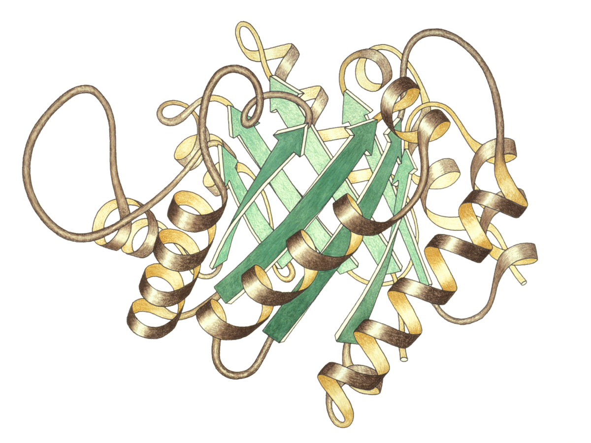
Triosephosphate isomerase (TIM) is the “perfect enzyme”. It is involved in glucose metabolism, and is present in high concentrations in muscle tissue, where it acts to generate ATP rapidly.

In glucose metabolism, glucose is broken down to the two substrates: glyceraldehyde-3-phosphate (G3P) and dihydroxyacetone phosphate (DHAP). This is an energetically unfavourable process, as it is inefficient and consumes ATP.

G3P is then converted to pyruvate, producing 2 ATP molecules and 1 NADH for each 3 carbon unit. The rate of this reaction, however, depends on the G3P concentration.

Through an equilibrium between G3P and DHAP in muscle tissues, TIM efficiently catalyses storage in a diffusion controlled conversion. This allows twenty-three times the amount of energy precursor than usual to be stored.

Triose Phosphate Isomerase
Equilibrium

The equilibrium functions at a rate determined by which the substrates can diffuse through the reaction medium to get to the enzyme.

TIM is so perfect that when artificially mutated, it mutates back to perfection, with a catalytic number of 10^8 – 10^9/M/s.

The mechanism for the reaction catalysed by TIM involves the formation of an enediol intermediate. The catalytic residues glutamate and histidine are involved in general acid-base catalysis.

General acid-base catalysis is involved in a majority of enzymatic reactions, wherein the side chains of various amino acids act as general acids or general bases. General acid-base catalysis involves a molecules besides water that acts as a proton donor or acceptor during the enzymatic reaction. It facilitates a reaction by stabilising the charges in the intermediate state, through the use of an acid or base.

Nucleophilic and electrophilic groups are activated as a result of the acid/base, and causes the reaction to proceed.

An example of acid-base catalysis is peptide hydrolysis by chymotrypsin.

Chymotrypsin - Wikipedia
Chymotrypsin

Chymotrypsin is involved in cleavage of amide bonds in peptides. It hydrolyses the peptide bond which connects the carboxyl group of one amino acid to the amino group of another. Key parts of the enzyme include serine, aspartic acid and histidine residues. The enzymatic reaction occurs in a step-wise process, generating a catalytic triad to improve the nucleophilic properties of serine and water.

Chymotrypsin uses a histidine residue and aspartic acid as a base catalyst. The histidine-aspartate deprotonate serine to increase its nuclephilicity. The serine then attacks the substrate’s carbonyl carbon, forming a tetrahedral intermediate. Then, an acyl group bounds to an intermediate, forming an acyl-enzyme intermediate. One product diffuses away at this time.

In carbonic anhydrase, the histidine residue helps the removal of hydrogen ion from the water molecule to generate OH- and strengthen its nucleophilic property. Once done, the water molecule attacks the acyl-enzyme, forming another tetrahedral intermediate, forming another product which diffuses away.

An oxyanion hole stabilises the tetrahedral intermediate anion formed during proteolysis and protects substrate’s negatively charged oxygen from water molecules. It stabilises the tetrahedral intermediate in chymotrypsin.

Enzymes can also attach to substrates “poised to strike”. Upon binding to a substrate, enzymes use binding energy to distort or strain the molecule on binding towards the transition state for the reaction, increasing energetic favourability.

Enzymes also work through binding in a reactive conformation. For example, phenylalanine ammonia lyase catalyses the the elimination of ammonia, and conversion of phenylalanine to cinnamic acid; putting phenylalaline amino acid into the food cycle. This reaction occurs in an “anti-configuration” environment as the hydrogen and amino group are lost, and henceforth biases the reaction.

Phenylalanine Ammonia-Lyase - an overview | ScienceDirect Topics
Penylalanine Ammonia Lyase Reaction

Genetics: RNA Translation

Translation is the process of translating the sequence of mRNA molecule to a sequence of amino acids during protein synthesis.

RNA translation has the following components:

  • mRNA to carry genetic information in the form of a codon.
  • tRNA to decipher the mRNA code and deliver specific amino acids to the ribosome.
  • rRNA to associate with a set of proteins to form a ribosome.
  • Ribosome to act as protein synthesis machinery.

There are four phases of translation:

  1. Charging of tRNA.
  2. Initiation.
  3. Elongation.
  4. Termination.

Genetics: RNA Transcription

Bacterial genes are often found in an operon.

Operons | BioNinja
Operon

An operon is essentially an assembly line of regulatory DNA genes, controlled by a region called the ‘promoter’. The regulatory DNA sequences act as binding sites for regulatory proteins, that promote or inhibit transcription.

Operons are quite efficient, however, if a singular mutation occurs at any point on the operon, the entire polycistronic pathway can be impacted. Operons can also struggle to take advantage of environmental changes; all genes are activated when the promoter is active.

The lac operon (article) | Gene regulation | Khan Academy
The lac Operon

A common example of an operon is the lac operon.

The lactose, or lac, operon is most commonly found in the bacterium Escherichia coli (E. coli). The lac operon refers to a cluster of three structural genes that each encode for proteins involved in lactose metabolism. These genes are ‘lacA’, ‘lacY’ and ‘lacZ’. LacZ and lacY are essential for the utilisation of lactose by E. coli.

The lac operon is a negative, inducible system. When no lactose is present, a repressor binds to the operator – preventing transcription. In the presence of lactose, allolactose binds and inactivates the lac repressor. This allows RNA polymerase to bind to the lac operon – enabling transcription.

It is important to note that a repressor binds to the operator, which prevents RNA polymerase from binding and transcription occurring. This is negative control.

An activator encourages polymerase to bind to the promoter. This is positive control.

The lacA gene encodes for lactose transacetylase; an enzyme that transfers an acetyl group from acetyl-CoA to galactosides.

The lacY gene encodes for lactose permease; a transmembrane protein that facilitates the movement of lactose across the phospholipid bilayers that surround all cells and organelles via active transport. When glucose is present, lactose permease is not produced – hence, lactose cannot be transported into the cell.

The lacZ gene encodes for β-galactosidase; a bacterial enzyme that catalyses the breakdown of lactose into its component simple monosaccharides, glucose and galactose. The synthesis of β-galactosidase is activated when glucose levels are low, and lactose is present. When glucose is low, β-galactosidase and lactose fit together. Once together, a change in conformation of the enzyme occurs. This new conformation causes bond strain between the monosaccharides, until eventually the bond breaks, and glucose and galactose dissociate from the enzyme to provide energy to the bacterial system. β-galactosidase synthesis stops when glucose levels are sufficient.

Lactose permease actively transports lactose into the cell. Following this, β-galactosidase breaks down the lactose into its components galactose and glucose. β-galactosidase also converts lactose into allolactose, then converts the allolactose into galactose and glucose.

Catabolite repression regulates the lac operon via positive control. It is the process of glucose repression. There is an inverse relationship between glucose and cyclic-AMP (cAMP); when cellular glucose levels are high, cAMP is low, and vice versa. When cAMP is present, a catabolite activator protein (CAP) binds to the lac operon promoter, facilitating the binding or RNA polymerase to the promoter, leading to enhanced transcription of the operon’s genes.

The lac operon (article) | Gene regulation | Khan Academy
The lac Operon

Another common operon example is the tryptophan, or trp, operon. The trp operon is an example for negative repressible transcription.

When tryptophan is low, the inactive regulator protein (repressor) does not bind to the operator, enabling transcription. However, when tryptophan is high, the repressor and tryptophan bind together, then bind to the operator. This prevents transcription from occurring.

Attenuation is a mechanism for reducing expression of the trp operon when levels of tryptophan are high. Rather than blocking initiation of transcription, attenuation prevents completion of transcription. The attenuation of the trp operon works through a mechanism that depends on coupling (the translation of an mRNA that is still in the process of being transcribed).

The trp RNA is able to form a hairpin. When sections 1 and 2 pair, and 3 and 4 align, transcription is terminated. However, when 2 and 3 bind, transcription still occurs. This determines which regions pair up.

The trp operon (article) | Gene regulation | Khan Academy
Low Tryptophan

When tryptophan levels are low, the ribosome stalls at the trp codons in region 1. Region 2 then is not covered by the ribosome, where region 3 is transcribed. When region 3 is transcribed, it pairs with region 2 – the attenuator never forms and transcription continues.

The trp operon (article) | Gene regulation | Khan Academy
High Tryptophan

When tryptophan levels are high, RNA polymerase begins transcribing DNA – producing region 1 of the 5′ UTR. A ribosome binds to the 5′ end of the 5′ UTR, and translates region 1 while region 2 is being transcribed.

RNA polymerase transcribes region 3. The ribosome does not stall at the trp codons, because tryptophan is abundant.

The ribosome covers part of region 2, preventing pairing with region 3. Region 4 is transcribed and pairs with region 3, producing the attenuator that terminates transcription.