An operon is essentially an assembly line of regulatory DNA genes, controlled by a region called the ‘promoter’. The regulatory DNA sequences act as binding sites for regulatory proteins, that promote or inhibit transcription.
Operons are quite efficient, however, if a singular mutation occurs at any point on the operon, the entire polycistronic pathway can be impacted. Operons can also struggle to take advantage of environmental changes; all genes are activated when the promoter is active.
The lac Operon
A common example of an operon is the lac operon.
The lactose, or lac, operon is most commonly found in the bacterium Escherichia coli (E. coli). The lac operon refers to a cluster of three structural genes that each encode for proteins involved in lactose metabolism. These genes are ‘lacA’, ‘lacY’ and ‘lacZ’. LacZ and lacY are essential for the utilisation of lactose by E. coli.
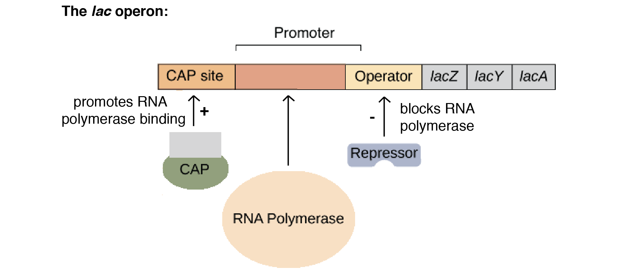
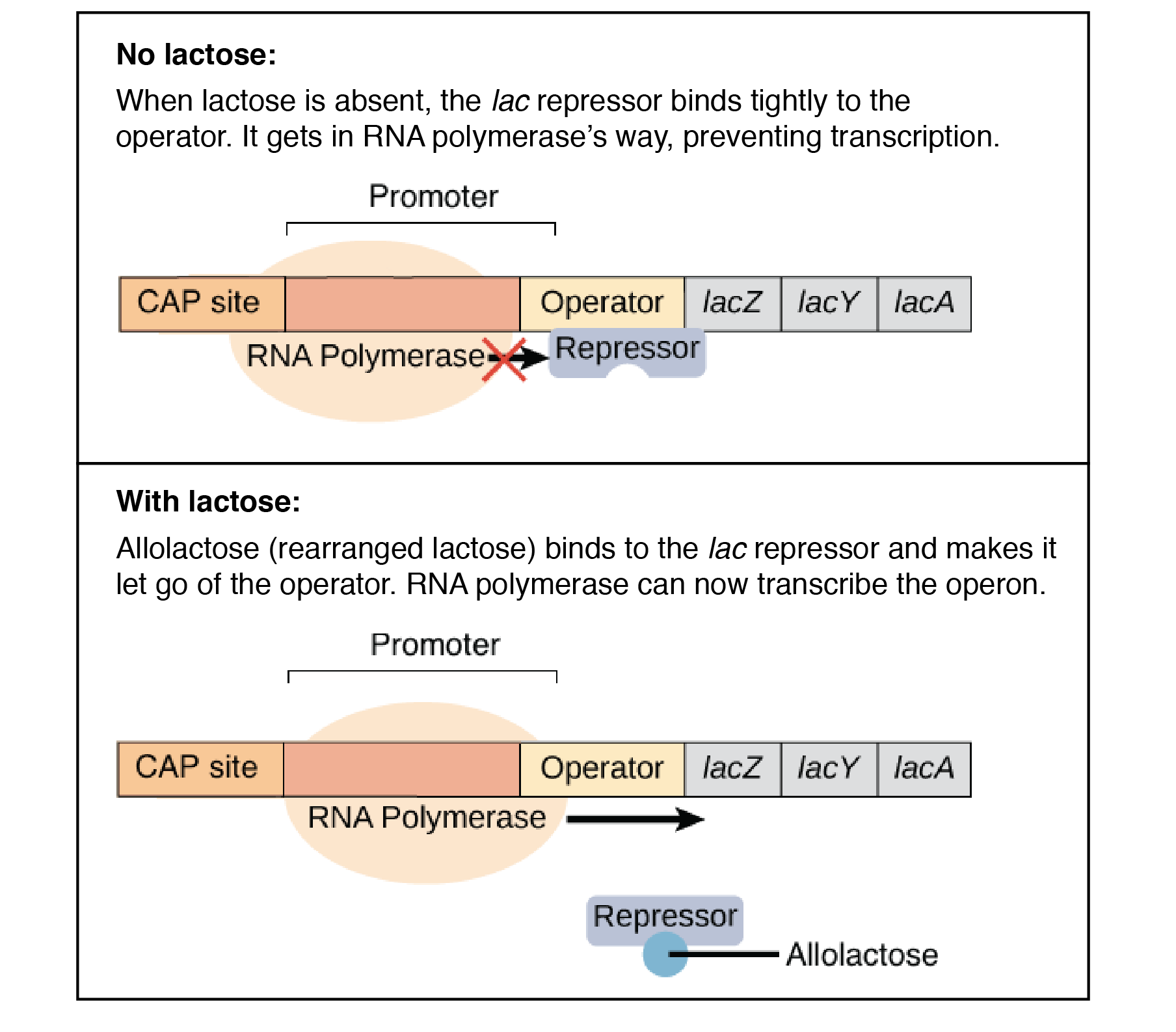
The lac operon is a negative, inducible system. When no lactose is present, a repressor binds to the operator – preventing transcription. In the presence of lactose, allolactose binds and inactivates the lac repressor. This allows RNA polymerase to bind to the lac operon – enabling transcription.
It is important to note that a repressor binds to the operator, which prevents RNA polymerase from binding and transcription occurring. This is negative control.
An activator encourages polymerase to bind to the promoter. This is positive control.
The lacA gene encodes for lactose transacetylase; an enzyme that transfers an acetyl group from acetyl-CoA to galactosides.
The lacY gene encodes for lactose permease; a transmembrane protein that facilitates the movement of lactose across the phospholipid bilayers that surround all cells and organelles via active transport. When glucose is present, lactose permease is not produced – hence, lactose cannot be transported into the cell.
The lacZ gene encodes for β-galactosidase; a bacterial enzyme that catalyses the breakdown of lactose into its component simple monosaccharides, glucose and galactose. The synthesis of β-galactosidase is activated when glucose levels are low, and lactose is present. When glucose is low, β-galactosidase and lactose fit together. Once together, a change in conformation of the enzyme occurs. This new conformation causes bond strain between the monosaccharides, until eventually the bond breaks, and glucose and galactose dissociate from the enzyme to provide energy to the bacterial system. β-galactosidase synthesis stops when glucose levels are sufficient.
Lactose permease actively transports lactose into the cell. Following this, β-galactosidase breaks down the lactose into its components galactose and glucose. β-galactosidase also converts lactose into allolactose, then converts the allolactose into galactose and glucose.
Catabolite repression regulates the lac operon via positive control. It is the process of glucose repression. There is an inverse relationship between glucose and cyclic-AMP (cAMP); when cellular glucose levels are high, cAMP is low, and vice versa. When cAMP is present, a catabolite activator protein (CAP) binds to the lac operon promoter, facilitating the binding or RNA polymerase to the promoter, leading to enhanced transcription of the operon’s genes.
The lac Operon
Another common operon example is the tryptophan, or trp, operon. The trp operon is an example for negative repressible transcription.
When tryptophan is low, the inactive regulator protein (repressor) does not bind to the operator, enabling transcription. However, when tryptophan is high, the repressor and tryptophan bind together, then bind to the operator. This prevents transcription from occurring.
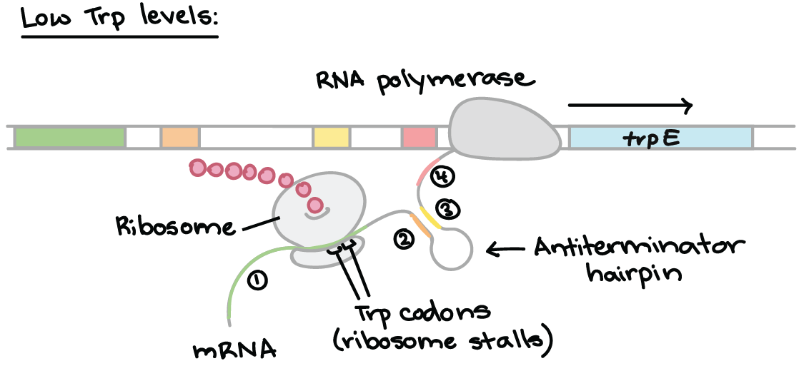
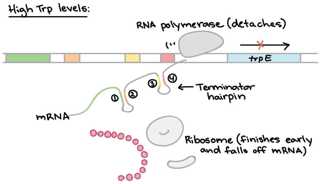
Attenuation is a mechanism for reducing expression of the trp operon when levels of tryptophan are high. Rather than blocking initiation of transcription, attenuation prevents completion of transcription. The attenuation of the trp operon works through a mechanism that depends on coupling (the translation of an mRNA that is still in the process of being transcribed).
The trp RNA is able to form a hairpin. When sections 1 and 2 pair, and 3 and 4 align, transcription is terminated. However, when 2 and 3 bind, transcription still occurs. This determines which regions pair up.
Low Tryptophan
When tryptophan levels are low, the ribosome stalls at the trp codons in region 1. Region 2 then is not covered by the ribosome, where region 3 is transcribed. When region 3 is transcribed, it pairs with region 2 – the attenuator never forms and transcription continues.
High Tryptophan
When tryptophan levels are high, RNA polymerase begins transcribing DNA – producing region 1 of the 5′ UTR. A ribosome binds to the 5′ end of the 5′ UTR, and translates region 1 while region 2 is being transcribed.
RNA polymerase transcribes region 3. The ribosome does not stall at the trp codons, because tryptophan is abundant.
The ribosome covers part of region 2, preventing pairing with region 3. Region 4 is transcribed and pairs with region 3, producing the attenuator that terminates transcription.
The definition of a gene is “the entire nucleic acid sequence that is necessary for the synthesis of a functional gene product (polypeptide or RNA)”.
Prokaryotic vs Eukaryotic Gene Structure
The gene structure of prokaryotes are essentially the “blueprint” for the bigger picture; the overall genome dictates what the prokaryote will look like and how it will function.
The genetic content of a prokaryote includes:
A circular chromosome.
A circular plasmid,
A nucleoid,
Genes organised as operons, polycistronic transcription.
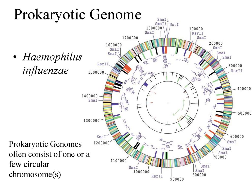
Prokaryotic genomes are part of one large circular DNA molecule.
Prokaryotic Genome
The bacterial genome includes one origin of replication. Just one origin of replication is required to initiate DNA replication – removing this origin would mean there is no propagation of DNA.
Most bacterial species contain circular chromosomal DNA, usually a few million base pair long. A few thousand different genes are interspersed throughout the genome; intergenic regions are regions that are not genes.
Genes are organised into operons and transcribed as a polycistronic mRNA. An operon consists of a set of genes encoding proteins participating in the same metabolic pathway.
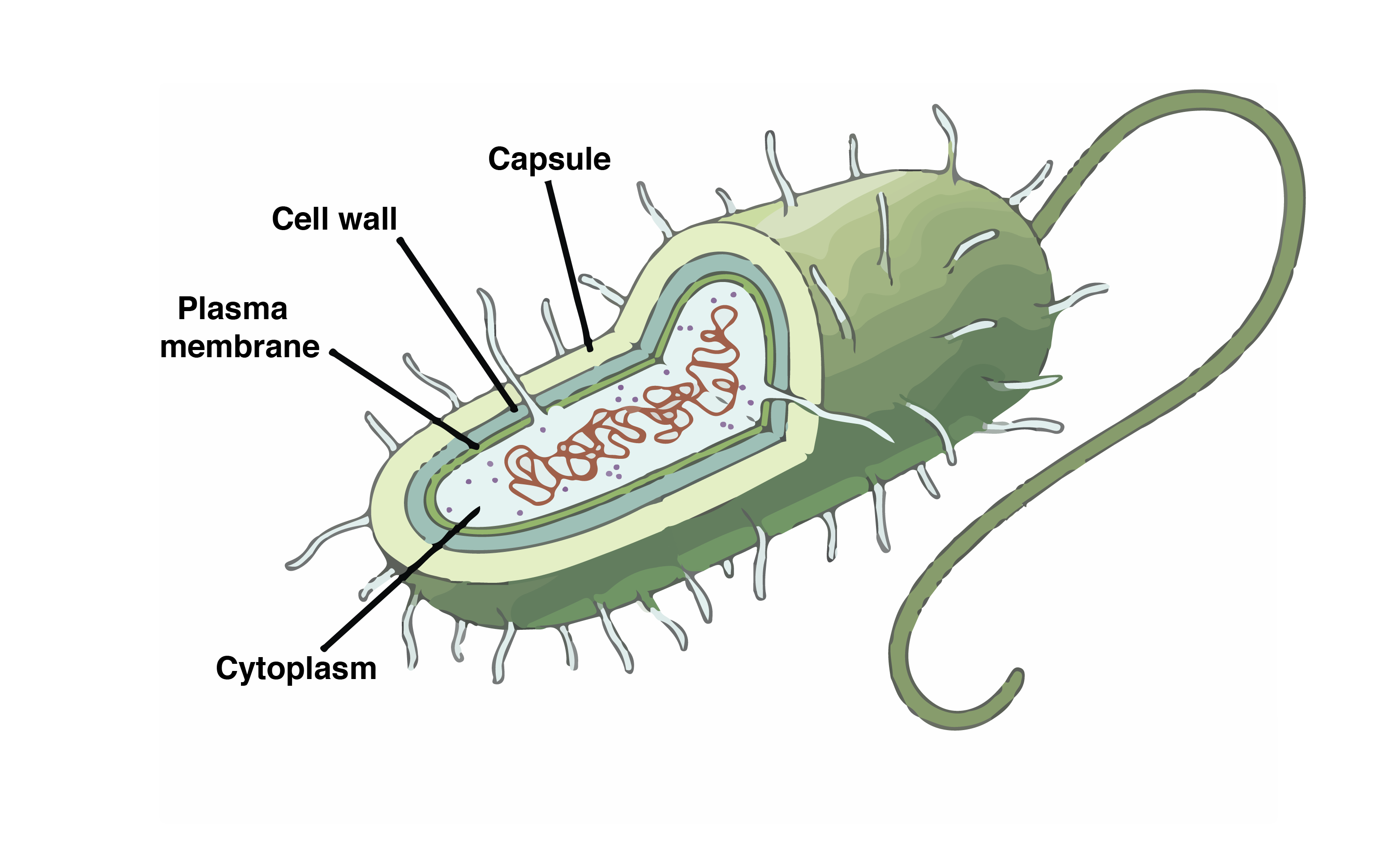
Prokaryotic Cell
Prokaryotes lack cell organisation. Unlike eurkaryotes, DNA is not complexed with histones. There is a relatively lesser amount of DNA in prokaryotes than in eukaryotes.
Most prokaryotes contain two types of DNA: bacterial chromosomes provide the main source of information, whereas plasmids are non-essential. Plasmids replicate independently, contain fewer genes, are not essential for survival, and can integrate with the chromosome.
Genes from plasmids can integrate into chromosomes. This means you can change bacteria from non-pathogenic to pathogenic, by adding plasmids with particular virulence factors. Information henceforth flows between plasmids and bacteria.
Prokaryotes multiply via binary fission; asexual reproduction. This means prokaryotes can replicate at an extremely rapid pace – for example, E. coli can replicate every 20 minutes.
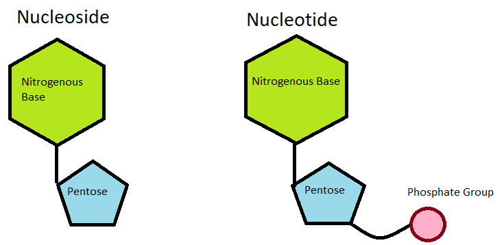
DNA (deoxyribonucleic acid) is the hereditary material found in almost all organisms. The function of DNA is determined by its structure.
The information of DNA is stored as a code made up of for chemical bases: adenine (A) and guanine (G) (the purines), and cytosine (C) and thymine (T) (the pyrimidines). Each of these bases make up the important DNA monomer unit, the nucleotide: which consists of a phosphate, pentose sugar and base.
Nucleoside vs Nucleotide
A common phrase people hear about DNA is that it is a “double helix”. This refers to the fact that DNA is made up of two complementary strands that are tightly wound together. DNA is twisted in this way due to hydrogen bonds forming between the bases (C-G, A-T), and ring polarisation of said bases. Van der Waals forces stabilise this twisting, via the sum of Van der Waals radii (3.4Å), enabling DNA to adopt the lowest energy state possible.
Separating the two strands of DNA, however, causes supercoiling. The term ‘supercoiling’ is an expression of the strain on that strand.
Supercoiling of DNA is an important biological process, that is regulated by topoisomerases and gyrases (specific DNA enzymes).
Topoisomerase and Gyrase Supercoiling a DNA Monomer
By supercoiling DNA, it can be easily compacted and utilised in further processes, such as DNA replication or transcription. By tightly wounding DNA, large amounts of it can be packed into the nucleus. This allows DNA to be safely stored but remain easily accessible.
A simple way to imagine this is by picturing an elastic band.
Elastic Band Supercoiling
By twisting and rolling the elastic band between your finger and thumb, the band shrivels and tightens, and becomes much smaller; compact. Imagine we have a small box we need to fill with these bands – it would be much easier to fill the box with compacted elastic than the original, large elastic. Even though it has changed shape, it is still an elastic band. It still provides the same information – it is just smaller. That is what DNA supercoiling is doing in the nucleus.
In a human cell, approximately six feet of DNA must be packaged into a nucleus with a diameter less than a human hair. To do this, nucleosomes are used.
Nucleosomes are the basic packing unit of eukaryotic DNA.
Nucleosome Structure
Each nucleosome is an octamer (polymer of eight molecules) of two copies of each of the nucleosomal histones, H2A, H2B, H3 and H4. 147 base pairs of DNA are wrapped almost twice around the histone octamer. Histone H1 binds outside of the nucleosome. Any non-histone proteins form a chromatin scaffold.
The nucleosomes are then arranged like beads on a string. They are repeatedly folded in on themselves to form a chromosome; a DNA molecule with genetic material of an organism.