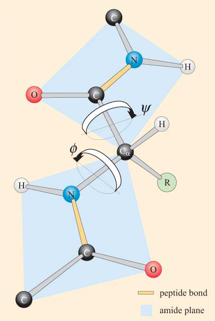
The peptide bond has a partial double bond character, caused by significant delocalisation of the lone pair of electrons on the nitrogen atom. This partial bond leads to a large dipole moment, and renders the amide group planar. There is a barrier of 88 kJ/mol between the carbon and nitrogen to keep the amide group planar. This rigidity of the peptide bond reduces the degrees of freedom of the polypeptide during folding.
The peptide bonds in the protein are usually in the trans configuration. The ‘R’ groups try to get “as far as well possible from each other”. Steric hindrance between the functional groups and the alpha-carbon atom will be greater in this cis formation.
Trans vs Cis Configuration
However, cis forms can occur in peptide bonds that precede a proline residue. In such cases, the cis form is more stable than usual, since the proline side-chain offers less of a hindrance.
Trans vs Cis Proline Configurations
The cyclic nature of proline means that both cis and trans configurations have more equivalent energies, as steric clashes occur in both. Thus, proline is found in the cis configuration more frequently than other amino acids.
In contrast, whilst rotation is not permitted about the peptide bonds, there is potential for rotation around the Cα–N and Cα–C bonds. This freedom of rotation about two bonds of each amino acid allows proteins to fold in many different ways. The rotations about these bonds can be specified by dihedral angles (also known as torsion angles).
The angle of rotation about the Cα–N bond is called phi (φ). The angle of rotation about the Cα–C bond is called psi (ψ).
The φ and ψ angles define the amount of rotation about the peptide bond, and determine the path of the polypeptide chain.
Phi and Psi Angles
Due to steric restraints, not all values of φ and ψ are available. The allowed values can be visualised on a two-dimensional plot called the Ramachandran plot.
The Ramachandran plot allows us to identify the sterically favourable conformations according to the following criteria:
Where there is no conflict between the van der Waals radii of non-bonding atoms, a conformation is ‘allowed’.
Conformations requiring interatomic distances at the limit which is permissible are defined as ‘outer limit’ conformations.
Theoretical conformations that require any two non-bonding atoms to be closer to each other than their van der Waals radii allow are sterically ‘forbidden’.
Ramachandran Plot
Three-quarters of the possible (φ, ψ) combinations are excluded simply by local steric clashes. Steric exclusion, the fact that two atoms cannot be in the same place at the same time, can be a powerful organizing principle.
The rigidity of the peptide unit and the restricted set of allowed φ and ψ angles limits the number of structures accessible to the unfolded form sufficiently to allow protein folding to occur.
Glycine and proline have much greater areas of conformational space available than other amino acids.
Glycine has greater conformational freedom due to its singular proton in its side-chain, making it less sterically hindered.
Trans-Glycine Plot
Proline has a unique, fused-ring structure, with no proton attached to its alpha-carbon.
Penicillium is a genus of fungi that plays a vital role in the pharmaceutical and medicinal industries. In 1929, Alexander Fleming, a bacteriologist in London, published a paper detailing an isolated Penicillium mould. He named the active agent of this mould ‘penicillin’, and determined that penicillin has an anti-bacterial effect on multiple pathogens.
Although Fleming is credited with the discovery of penicillin, it is likely that penicillin has been in use since prehistoric times. Ancient Egypt and Ancient Greece, famously, each used an enormous assortment of such moulds for an arrangement of medicinal uses.
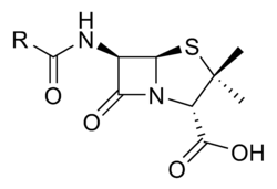
The versatile pharmacological effect of the fungi is due to their unique structure.
Penicillin General Structure
Penicillin comes in various forms, such as Penicillin G (benzylpenicillin) and Penicillin V (phenoxymethylpenicillin). Other β-lactam antibiotics include cephalosporins. The characteristic feature of all penicillins and cephalosporins is the 4-membered cyclic amide, or β-lactam ring. The ring has a high bond strain of 90° – making it a highly reactive molecule once the strain is released.
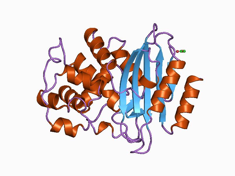
Penicillin kills bacteria through the inhibition of the enzyme DD-transpeptidase via the β-lactam ring.
DD-transpeptidases synthesise bacterial cell walls – by inhibiting this enzyme, new cell wall formation is also inhibited; the bacterial cell wall becomes vulnerable to external forces and subsequently dies.
DD-Transpeptidase
Penicillin compounds highly resemble D-Ala-D-Ala amides. Hence, when a penicillin binds to the DD-transpeptidases, the serine will attack the amide bond of the β-lactam group, and an ester will form. The new five-membered ring henceforth blocks further binding to the enzyme active site.
Penicillin vs D-Ala-D-Ala
The detection of this particular mechanism within the penicillin compounds has significantly changed the process of infectious disease treatment; bacterial diseases can be combatted. Modern medicine has been refined to reproduce the process within the Penicillium genus responsible for medicinal value. This has led to a large, industrial-level development of antibiotics.
Antibiotics are drugs that help stop infections caused by bacteria. For example, Penicillin G was the original penicillin ‘discovered’ by Fleming. It came into commercial use in 1942, and was famously used for treatment during WWII. Penicillin G antibiotics treat a wide variety of bacterial infections, including diphtheria, pneumonia, gas gangrene and tetanus. It is mostly administered via injection.
Biosynthesis of the β-lactam antibiotics begins with the biosynthesis of the common precursor amino acids: a homologue of glutamic acid, an L-cysteine and a D- valine. These amino acids link via peptide bonds. An isopenicillin N synthetase then catalyses the cyclisation of the Arnstein peptide through to give isopenicillin N via REDOX reaction.
After this, a peptidase cleaves the amide bond to produce 6-aminopenicillicacid (6-APA). The REDOX co-factors in this reaction, Fe2+ and O2, assist in this cleavage. Acylating the resulting nitrogen compound leads to the development of the penicillin antibiotic.
Cephalosporins are synthesised with cephalosporin synthetase. The continuing biosynthesis follows the same pathway as penicillins.
Penicillin G Synthesis
Unfortunately, with the use of antibiotics, comes the development of antibiotic resistant-bacteria. Causes of antibiotic resistance vary, however, are predominantly the result of over-prescription, over-use, and patients not finishing their course. Bacteria, such as E. coli, have evolved new mechanisms to overcome the inhibitory effects of β-lactamase inhibitors. There are several mechanisms bacteria use, however, a common one is drug inactivation or modification – some penicillin-resistant bacteria have developed the ability to enzymatically deactivate penicillin.
β-Lactamases are enzymes produced by some bacterial cell walls that have the ability to convert penicillin to the biologically inactive ‘penicilloic’, by adding an acetyl or phosphate group to the specific site on the antibiotic. This effectively disrupts the functioning of the antibiotic; causing the bacteria to be resistant to the β-lactam ring attack.
β-lactamase
This is a very effective mechanism, as not only do β-lactamases turn over substrate at ~2000/sec, the outer-membrane of the gram-negative, and peptidoglycan layer for the gram-positive bacteria, filter the penicillin getting through to the penicillin binding protein. The net result of this, is that β-lactamases are a common form of antibiotic resistance.
Gram-Negative vs Gram-Positive Bacterial Cell Walls
This resistance has the potential to have dire effects on the health industry, and furthermore, the overall population’s health – as bacteria overcoming such inhibitors renders the preferred use of medicinal penicillin invalid.
Many strategies to fight bacterial resistance have been developed. β-lactamase inhibitors, such as clavulanate, have the ability to bind to penicillin-binding proteins and overcome antibiotic resistance in bacteria that secrete β-lactamase. To put simply, β-lactamase inhibitors are able to inhibit β-lactamase. By combining β-lactamase inhibitors with penicillin in the form of a drug, β-lactamase can be overcome, and the challenges of antibiotic resistance can be more readily tackled. Further preventative measures, such as prescribing antibiotics only when necessary, limiting the use of antibiotics, and education patients to completely finish their course, can also help prevent the development of antibiotic resistance.
When penicillin resistance first came about, the initial response was to utilise cephalosporins. The reason why cephalosporins worked is related to their double bond in their six-membered ring.
Cephalosporin Structure
When the β-lactam antibiotics react with the β-lactamase, an ester is formed, and the the electrons of the cephalosporin core nitrogen are delocalised over the double bond to create a weak amine; the base catalysis of the ester hydrolysis is reduced. However, resistance was quickly developed against this.
Methicillin and flucloxacillin were the next to be developed.
Methicillin
Flucloxacillin
Methicillin and flucloxacillin have bulky, hydrophobic substituent, at the top left position of the β-lactam. This substituent can fit into the binding pocket of the penicillin binding proteins better than the binding pocket of the β-lactamase enzymes. This is because the β-lactamase enzymes are less hydrophobic and allow water through to hydrolyse esters. The penicillin binding proteins petition more effectively. However, further β-lactamase mutation then occurred – leading to more resilience.
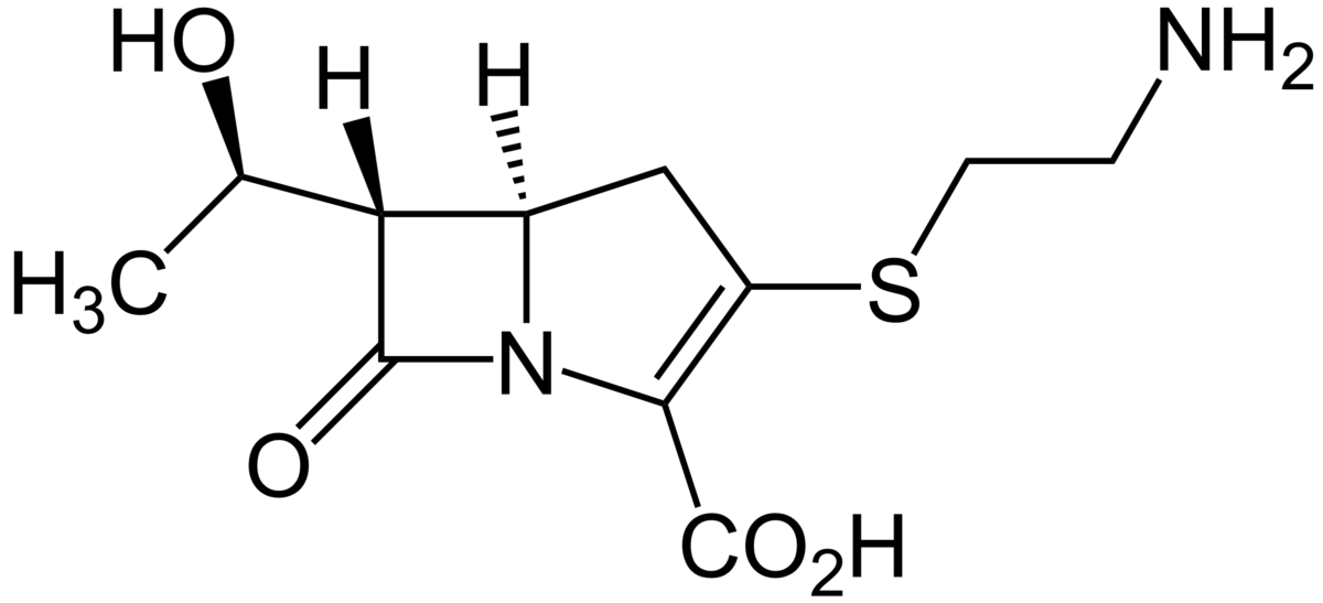
Thienamycin was then developed.
Thienamycin
Thienamycin is the 5th generation antibiotic. It functions similarly to the cephalosporin. Thienamycin inhibit the β-lactamase enzyme by forming an ester intermediate, but are less resistant to hydrolysis. The problem with thienamycin is that the active groups involved destroy the antibiotic itself. Henceforth, thienamycin was modified.
The modified products of thienamycin were imipenem and cilastatin.
Imipenem (top) Cilastatin (bottom)
By tying up the top-left amino group of the thienamycin, the compounds become stable. However, imipenem is removed in the kidneys of patients. It is recognised by the dehydropeptidase enzyme that removes dehydroamino acids. Cilastatin was henceforth developed to inhibit the dehydropeptidase. Both drugs were then combined to inhibit the β-lactamase. However, an issue with combining these drugs is that their effectiveness varies depending on their bioavailability across the gastro-intestinal tract. The compounds have different polarity and distribution across the body, and henceforth are rarely administered as an antibiotic.
Sulbactam is the sixth generation antibiotic to have been developed.
Sulbactam
Sulbactam has no side-chain in the top-left position of the β-lactam ring. Sulbactam works because of its charged substituent; a good leaving group. Base hydrolysis of the ester does not occur. Sulbactam strays from equilibirum between an amine and imine, making it more effective. However, it does not bind to the penicillin binding proteins due to its lack of substituent – and must be combined with another β-lactam antibiotic.
Unasyn is the combination of sulbactam and ampicillin. However, like before, these compounds vary drastically in their polarity and bioavailability – hence, sultamicillin was developed.
Sultamicillin
Sultamicillin effectively ties the two products together, allowing the compounds to transfer into the bloodstream at relatively the same rate and concentration.
Clavulanic acid is the seventh generation response to bacteria.
Clavulanic Acid
Clavulanic acid reacts selectively with the β-lactamase enzymes. It opens the β-lactam ring to form an ester intermediate, which can undergo base catalysed hydrolysis to cleave and release the material. Clavulanic acid has an inolate leaving group, which generates an imine. Following this, an irreversible reaction occurs, wherein an inhibitor enzyme attaches. It is a “suicide substrate, irreversible inhibitor”.
Clavulanic acid must be combined with an antibiotic to work effectively. Augmentin is the combination of clavulanic and amoxycillin.
Augmentin Tablets
Drugs of last resort are vancomycin (used by injection) and linezolid (used for long-term infections). These drugs have serious side-effects and are quite unstable, hence, are only used in hospital circumstances.
Enzymes achieve catalysis due to the active site cleft present in proteins.
Protein Active Site
The most important aspect of catalysis is species proximity – the closer the species are to one another, the better.
The rate of reaction of two species can be given by the equation:
rate = k[A].[B]
Typically, reagents without an enzyme have a nanomolar concentration and small rate. When together, however, the enzyme protein brings about the correct orientation of the species for a productive collision; bringing about a higher concentration and larger rate.
Enzymes function perfectly at the “diffusion control limit”. This is what enables enzymes to provide simultaneous acid-base catalysis.
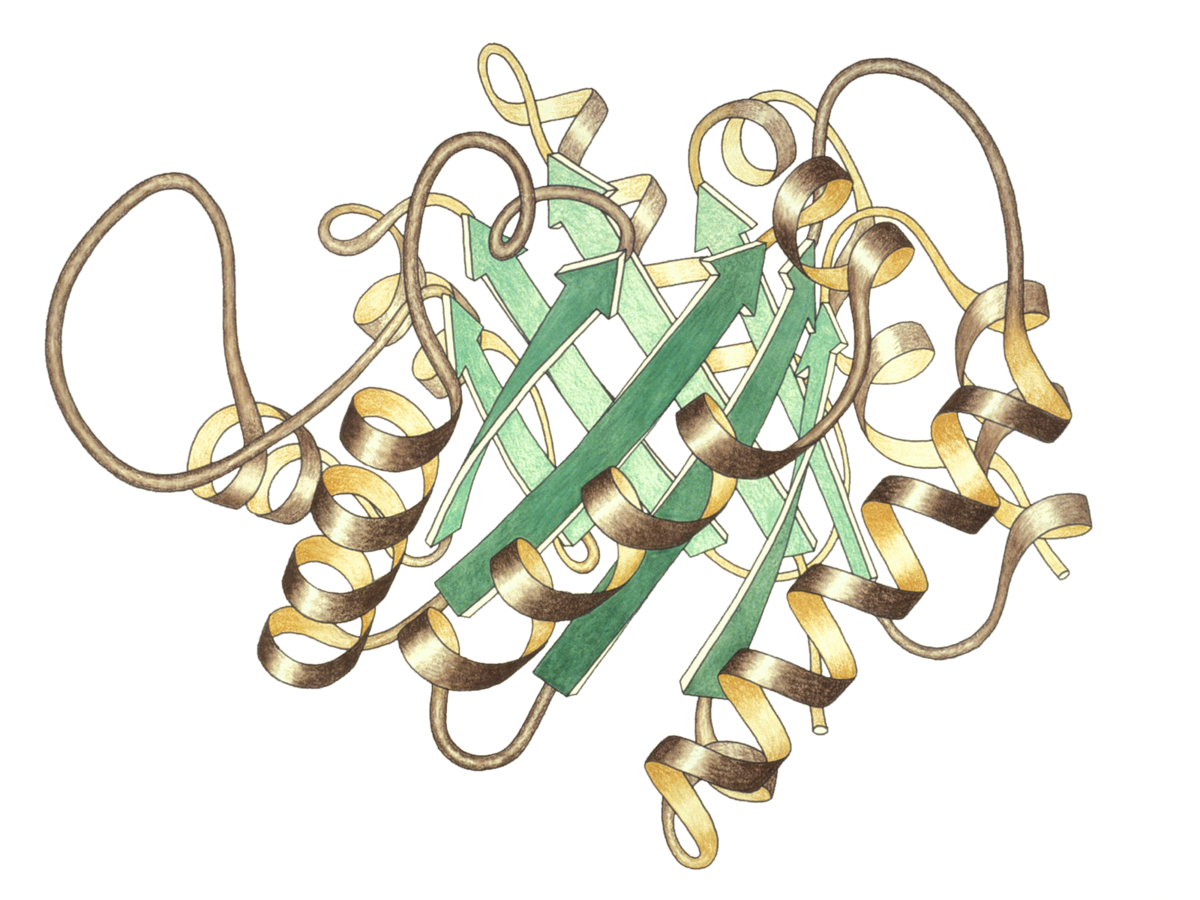
Triosephosphate Isomerase
Triosephosphate isomerase (TIM) is the “perfect enzyme”. It is involved in glucose metabolism, and is present in high concentrations in muscle tissue, where it acts to generate ATP rapidly.
In glucose metabolism, glucose is broken down to the two substrates: glyceraldehyde-3-phosphate (G3P) and dihydroxyacetone phosphate (DHAP). This is an energetically unfavourable process, as it is inefficient and consumes ATP.
G3P is then converted to pyruvate, producing 2 ATP molecules and 1 NADH for each 3 carbon unit. The rate of this reaction, however, depends on the G3P concentration.
Through an equilibrium between G3P and DHAP in muscle tissues, TIM efficiently catalyses storage in a diffusion controlled conversion. This allows twenty-three times the amount of energy precursor than usual to be stored.
Equilibrium
The equilibrium functions at a rate determined by which the substrates can diffuse through the reaction medium to get to the enzyme.
TIM is so perfect that when artificially mutated, it mutates back to perfection, with a catalytic number of 10^8 – 10^9/M/s.
The mechanism for the reaction catalysed by TIM involves the formation of an enediol intermediate. The catalytic residues glutamate and histidine are involved in general acid-base catalysis.
General acid-base catalysis is involved in a majority of enzymatic reactions, wherein the side chains of various amino acids act as general acids or general bases. General acid-base catalysis involves a molecules besides water that acts as a proton donor or acceptor during the enzymatic reaction. It facilitates a reaction by stabilising the charges in the intermediate state, through the use of an acid or base.
Nucleophilic and electrophilic groups are activated as a result of the acid/base, and causes the reaction to proceed.
An example of acid-base catalysis is peptide hydrolysis by chymotrypsin.
Chymotrypsin
Chymotrypsin is involved in cleavage of amide bonds in peptides. It hydrolyses the peptide bond which connects the carboxyl group of one amino acid to the amino group of another. Key parts of the enzyme include serine, aspartic acid and histidine residues. The enzymatic reaction occurs in a step-wise process, generating a catalytic triad to improve the nucleophilic properties of serine and water.
Chymotrypsin uses a histidine residue and aspartic acid as a base catalyst. The histidine-aspartate deprotonate serine to increase its nuclephilicity. The serine then attacks the substrate’s carbonyl carbon, forming a tetrahedral intermediate. Then, an acyl group bounds to an intermediate, forming an acyl-enzyme intermediate. One product diffuses away at this time.
In carbonic anhydrase, the histidine residue helps the removal of hydrogen ion from the water molecule to generate OH- and strengthen its nucleophilic property. Once done, the water molecule attacks the acyl-enzyme, forming another tetrahedral intermediate, forming another product which diffuses away.
An oxyanion hole stabilises the tetrahedral intermediate anion formed during proteolysis and protects substrate’s negatively charged oxygen from water molecules. It stabilises the tetrahedral intermediate in chymotrypsin.
Enzymes can also attach to substrates “poised to strike”. Upon binding to a substrate, enzymes use binding energy to distort or strain the molecule on binding towards the transition state for the reaction, increasing energetic favourability.
Enzymes also work through binding in a reactive conformation. For example, phenylalanine ammonia lyase catalyses the the elimination of ammonia, and conversion of phenylalanine to cinnamic acid; putting phenylalaline amino acid into the food cycle. This reaction occurs in an “anti-configuration” environment as the hydrogen and amino group are lost, and henceforth biases the reaction.
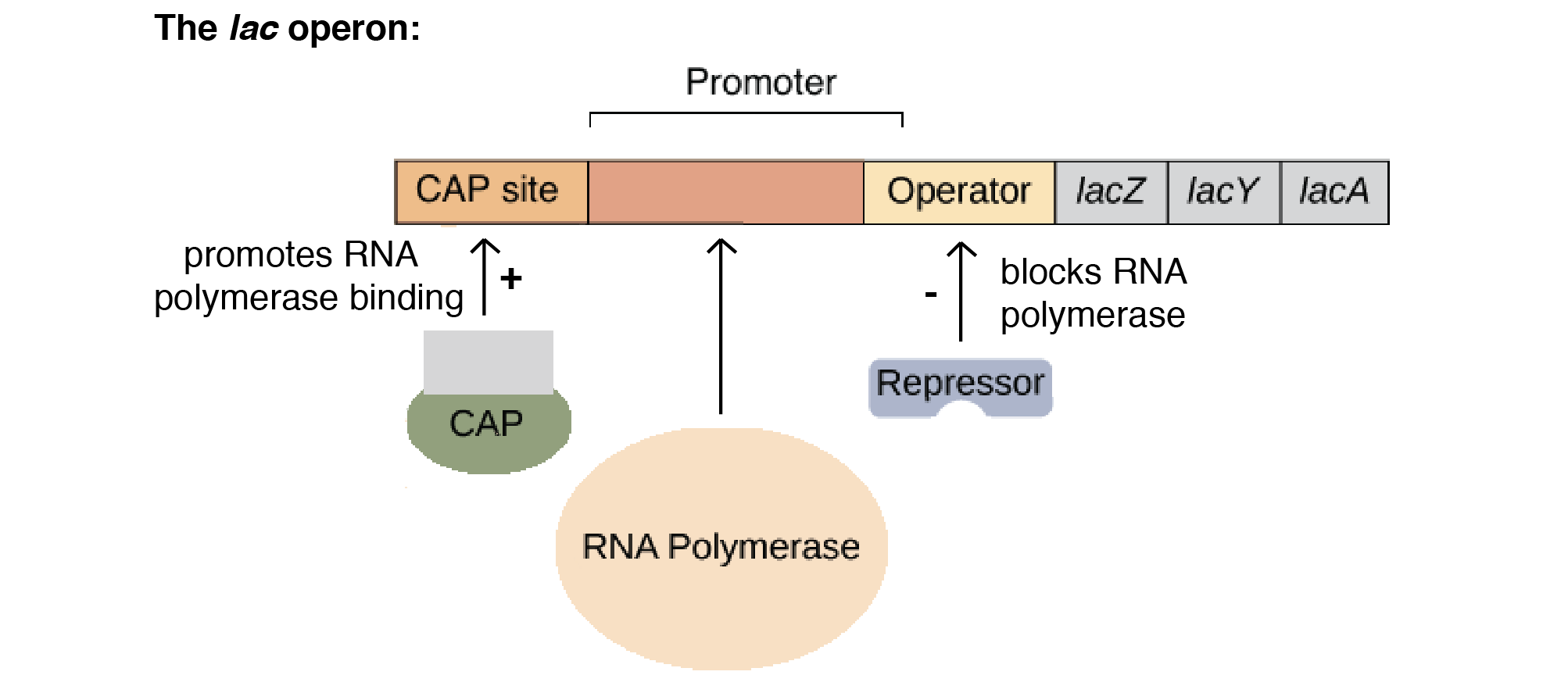
An operon is essentially an assembly line of regulatory DNA genes, controlled by a region called the ‘promoter’. The regulatory DNA sequences act as binding sites for regulatory proteins, that promote or inhibit transcription.
Operons are quite efficient, however, if a singular mutation occurs at any point on the operon, the entire polycistronic pathway can be impacted. Operons can also struggle to take advantage of environmental changes; all genes are activated when the promoter is active.
The lac Operon
A common example of an operon is the lac operon.
The lactose, or lac, operon is most commonly found in the bacterium Escherichia coli (E. coli). The lac operon refers to a cluster of three structural genes that each encode for proteins involved in lactose metabolism. These genes are ‘lacA’, ‘lacY’ and ‘lacZ’. LacZ and lacY are essential for the utilisation of lactose by E. coli.
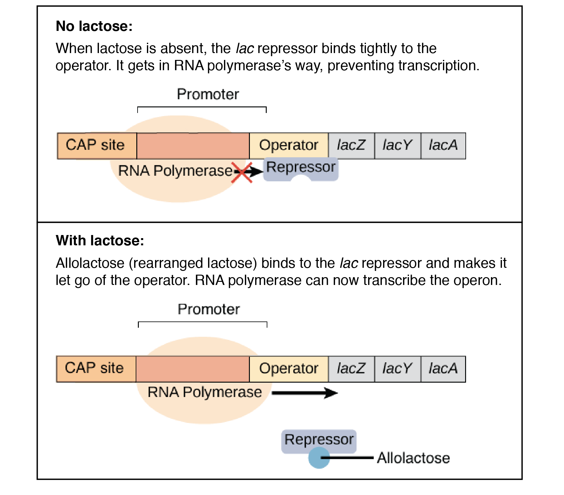
The lac operon is a negative, inducible system. When no lactose is present, a repressor binds to the operator – preventing transcription. In the presence of lactose, allolactose binds and inactivates the lac repressor. This allows RNA polymerase to bind to the lac operon – enabling transcription.
It is important to note that a repressor binds to the operator, which prevents RNA polymerase from binding and transcription occurring. This is negative control.
An activator encourages polymerase to bind to the promoter. This is positive control.
The lacA gene encodes for lactose transacetylase; an enzyme that transfers an acetyl group from acetyl-CoA to galactosides.
The lacY gene encodes for lactose permease; a transmembrane protein that facilitates the movement of lactose across the phospholipid bilayers that surround all cells and organelles via active transport. When glucose is present, lactose permease is not produced – hence, lactose cannot be transported into the cell.
The lacZ gene encodes for β-galactosidase; a bacterial enzyme that catalyses the breakdown of lactose into its component simple monosaccharides, glucose and galactose. The synthesis of β-galactosidase is activated when glucose levels are low, and lactose is present. When glucose is low, β-galactosidase and lactose fit together. Once together, a change in conformation of the enzyme occurs. This new conformation causes bond strain between the monosaccharides, until eventually the bond breaks, and glucose and galactose dissociate from the enzyme to provide energy to the bacterial system. β-galactosidase synthesis stops when glucose levels are sufficient.
Lactose permease actively transports lactose into the cell. Following this, β-galactosidase breaks down the lactose into its components galactose and glucose. β-galactosidase also converts lactose into allolactose, then converts the allolactose into galactose and glucose.
Catabolite repression regulates the lac operon via positive control. It is the process of glucose repression. There is an inverse relationship between glucose and cyclic-AMP (cAMP); when cellular glucose levels are high, cAMP is low, and vice versa. When cAMP is present, a catabolite activator protein (CAP) binds to the lac operon promoter, facilitating the binding or RNA polymerase to the promoter, leading to enhanced transcription of the operon’s genes.
The lac Operon
Another common operon example is the tryptophan, or trp, operon. The trp operon is an example for negative repressible transcription.
When tryptophan is low, the inactive regulator protein (repressor) does not bind to the operator, enabling transcription. However, when tryptophan is high, the repressor and tryptophan bind together, then bind to the operator. This prevents transcription from occurring.
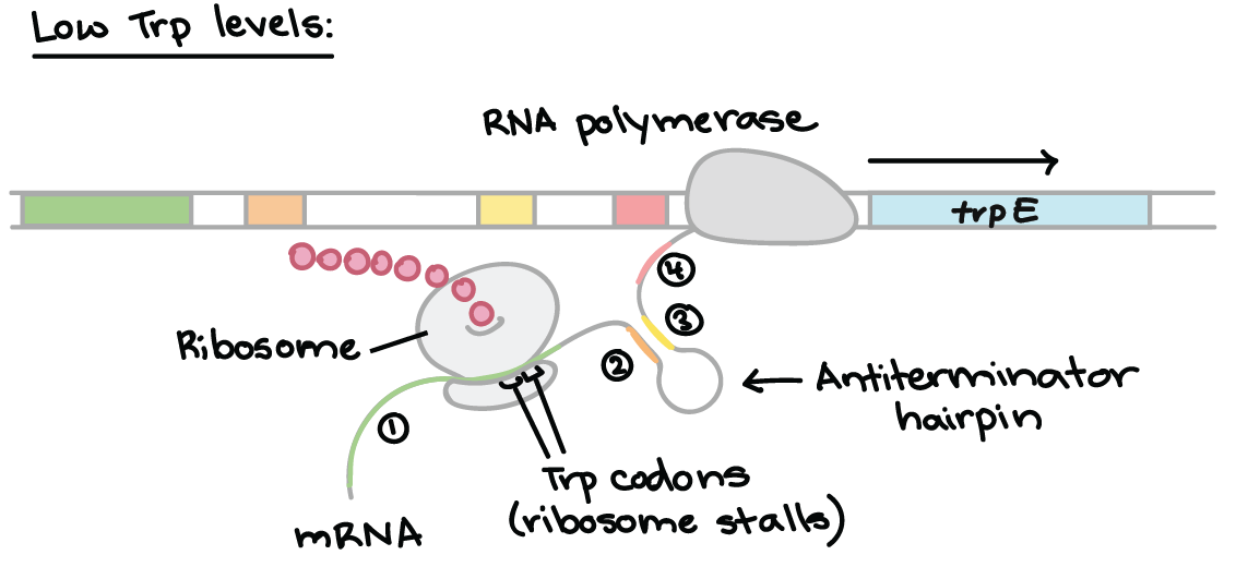
Attenuation is a mechanism for reducing expression of the trp operon when levels of tryptophan are high. Rather than blocking initiation of transcription, attenuation prevents completion of transcription. The attenuation of the trp operon works through a mechanism that depends on coupling (the translation of an mRNA that is still in the process of being transcribed).
The trp RNA is able to form a hairpin. When sections 1 and 2 pair, and 3 and 4 align, transcription is terminated. However, when 2 and 3 bind, transcription still occurs. This determines which regions pair up.
Low Tryptophan
When tryptophan levels are low, the ribosome stalls at the trp codons in region 1. Region 2 then is not covered by the ribosome, where region 3 is transcribed. When region 3 is transcribed, it pairs with region 2 – the attenuator never forms and transcription continues.
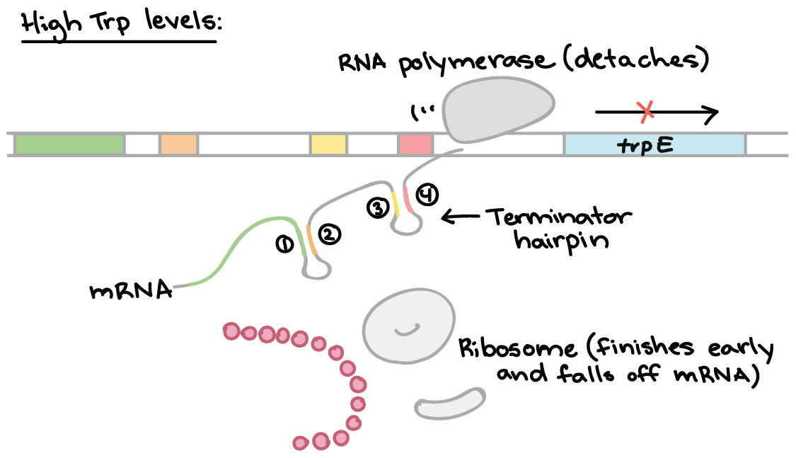
High Tryptophan
When tryptophan levels are high, RNA polymerase begins transcribing DNA – producing region 1 of the 5′ UTR. A ribosome binds to the 5′ end of the 5′ UTR, and translates region 1 while region 2 is being transcribed.
RNA polymerase transcribes region 3. The ribosome does not stall at the trp codons, because tryptophan is abundant.
The ribosome covers part of region 2, preventing pairing with region 3. Region 4 is transcribed and pairs with region 3, producing the attenuator that terminates transcription.
A common question those in science wonder is: “what is the difference between biochemistry and chemical biology?”.
Well, the difference is this: biochemistry typically focuses on the chemistry of biology, whereas chemical biology focuses on manipulating chemistry to solve biological problems.
Chemical biology can become complicated quite quickly, so it is important to understand and be comfortable with the basic biochemical processes within our body. Having the foundations of chemical biology down pact is valuable to a science student focusing on medicine, forensics, chemistry or biology. The foundations of chemical biology are also particularly handy for those wanting to sit the GAMSAT; the graduate medical entry examination that is held in Australia and the United Kingdom.
In this blog, we are going to discuss these major topics:
DNA (deoxyribonucleic acid) is the hereditary material found in almost all organisms. The function of DNA is determined by its structure.
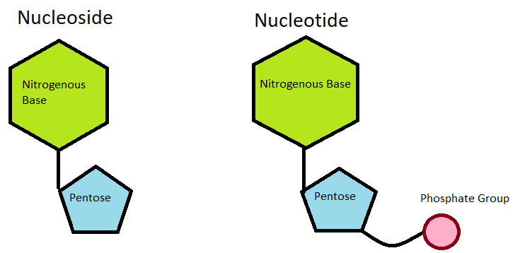
The information of DNA is stored as a code made up of for chemical bases: adenine (A) and guanine (G) (the purines), and cytosine (C) and thymine (T) (the pyrimidines). Each of these bases make up the important DNA monomer unit, the nucleotide: which consists of a phosphate, pentose sugar and base.
Nucleoside vs Nucleotide
A common phrase people hear about DNA is that it is a “double helix”. This refers to the fact that DNA is made up of two complementary strands that are tightly wound together. DNA is twisted in this way due to hydrogen bonds forming between the bases (C-G, A-T), and ring polarisation of said bases. Van der Waals forces stabilise this twisting, via the sum of Van der Waals radii (3.4Å), enabling DNA to adopt the lowest energy state possible.
Separating the two strands of DNA, however, causes supercoiling. The term ‘supercoiling’ is an expression of the strain on that strand.
Supercoiling of DNA is an important biological process, that is regulated by topoisomerases and gyrases (specific DNA enzymes).
Topoisomerase and Gyrase Supercoiling a DNA Monomer
By supercoiling DNA, it can be easily compacted and utilised in further processes, such as DNA replication or transcription. By tightly wounding DNA, large amounts of it can be packed into the nucleus. This allows DNA to be safely stored but remain easily accessible.
A simple way to imagine this is by picturing an elastic band.
Elastic Band Supercoiling
By twisting and rolling the elastic band between your finger and thumb, the band shrivels and tightens, and becomes much smaller; compact. Imagine we have a small box we need to fill with these bands – it would be much easier to fill the box with compacted elastic than the original, large elastic. Even though it has changed shape, it is still an elastic band. It still provides the same information – it is just smaller. That is what DNA supercoiling is doing in the nucleus.
In a human cell, approximately six feet of DNA must be packaged into a nucleus with a diameter less than a human hair. To do this, nucleosomes are used.
Nucleosomes are the basic packing unit of eukaryotic DNA.
Nucleosome Structure
Each nucleosome is an octamer (polymer of eight molecules) of two copies of each of the nucleosomal histones, H2A, H2B, H3 and H4. 147 base pairs of DNA are wrapped almost twice around the histone octamer. Histone H1 binds outside of the nucleosome. Any non-histone proteins form a chromatin scaffold.
The nucleosomes are then arranged like beads on a string. They are repeatedly folded in on themselves to form a chromosome; a DNA molecule with genetic material of an organism.